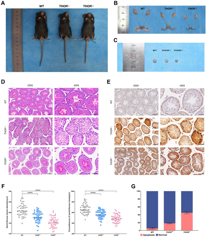
Figure 4.
THOR deficiency affects testicular development in male mice. (A) Photos of 8-week-old male WT and THOR-deficient mice. From left to right are WT, THOR−/−, THOR+/− mice. (B) Comparison of the reproductive systems of 8-week-old male WT and THOR-deficient mice. From left to right are WT, THOR+/−, THOR−/− mice. (C) Comparison of the testicular morphologies of 8-week-old WT and THOR-deficient mice. From left to right are WT, THOR+/−, THOR−/− mice. (D) H&E-stained tracheal sections of the testes of WT mice and THOR-deficient mice. In the testes of THOR-deficient mice, the seminiferous tubules had decreased diameters and were sparsely arranged and Sertoli and germ cells were arranged abnormally. (E) Immunohistochemical staining of the testes of WT and THOR-deficient mice. (F) Comparison of the diameters and circumferences of WT and THOR-deficient mouse seminiferous tubules. **** indicates p ≤ 0.0001 as determined by a t-test. The data are shown as the mean ± SD from two independent experiments. (G) The apoptotic rates of Sertoli cells in the seminiferous tubules of WT and THOR-deficient mice were determined by immunohistochemistry.

