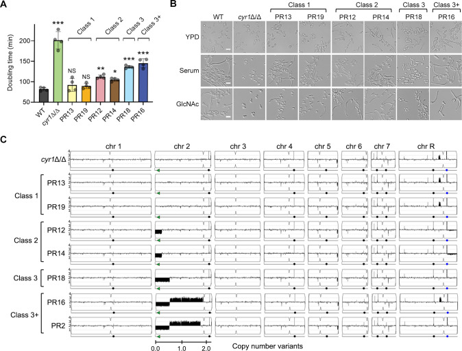
Fig 1. Genetic changes in chromosome 2 bypassed the need for cAMP to improve growth and hyphal induction in cyr1Δ/Δ PR mutants.
(A) Doubling times were measured in liquid YPD medium at 30°C. Shown is the mean ± SD (standard deviation) of 4 independent experiments. Statistical analysis was performed using one-way ANOVA with Dunnett’s multiple comparisons test comparing the strains with the wild-type control (WT); NS p > 0.05, * p < 0.05, ** p < 0.01, *** p < 0.001. The doubling time of the strains shown was significantly shorter when compared to the cyr1Δ/Δ background (p < 0.0001). (B) The strains indicated at the top were grown in the liquid medium indicated on the left, and then hyphal induction was assessed microscopically. Cells were grown in liquid medium containing 15% serum or 50 mM N-acetylglucosamine (GlcNAc) to induce hyphal growth. Cells were incubated at 37°C for 3 h and then photographed. Scale bar, 10 μm. (C) Copy number variation analysis based upon read depth across the genome. Copy number estimates scaled to genome ploidy (Y-axis) and chromosome location (X-axis) were plotted using YMAP [91]. Numbers and symbols below chromosomes indicate chromosomal position (Mb), BCY1 gene (green arrows), centromere locus (indentations in the chromosome cartoon), major repeat sequence position (black circles), and rDNA locus (blue circles, ChrR).

