
Figure 1.
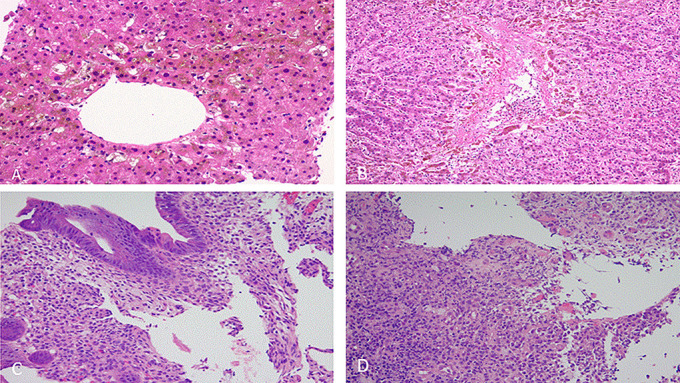
(A) Liver: canalicular cholestasis surrounding the central veins with associated bile duct injury. (B) Liver: zone 3 cholestasis and bile duct injury with patchy hepatocyte necrosis. Hematoxylin and eosin stain. (C) Sigmoid colon: surface epithelium with reactive changes, eosinophilic cytoplasm, and disorganized epithelial cell placement. Rare neutrophils are seen with the underlying lamina propria devoid of the normal glandular elements. (D) Sigmoid colon: ulcerated mucosa replaced with granulation tissue (hematoxylin and eosin stain).