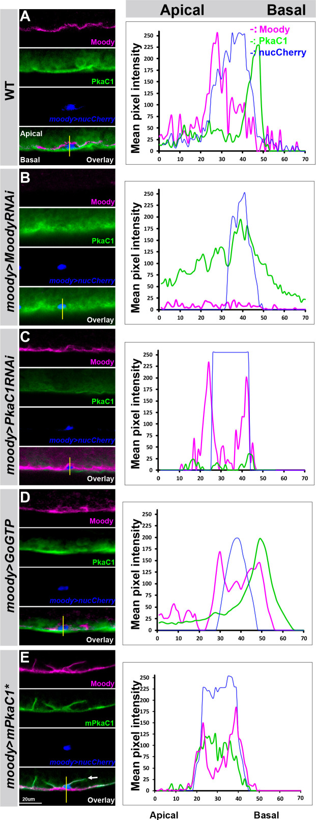
Figure 5. The Moody/protein kinase A (PKA) signaling pathway is polarized in subperineural glia (SPG).
The subcellular localization of the PKA catalytic subunit PkaC1 and Moody in SPG of third instar larvae in WT (A), Moody knockdown (moody>MoodyRNAi) (B), PkaC1 knockdown (moody>Pka-C1-RNAi), GPCR gain of function (moody>Go GTP), and PKA overexpression (moody>mPka-C1*). Antibody labeling of Moody (magenta), of Drosophila PkaC1 or mouse PkaC1 (green), and of SPG nuclei (moody>nucCherry; blue). (A–E) Lateral views of the CNS/hemolymph border, with CNS facing top. On the right column, line scans of fluorescence intensities for each channel along the apical–basal axis at the positions indicated. In WT (A), Moody localizes to the apical side and PkaC1 is enriched at the basal side of SPG. Under loss of Moody signaling (moody>MoodyRNAi) (B), PKA spreads throughout the cell and loses its basal localization. Moody loses its apical localization under reduced (C) or increased PKA activity (E). Under GPCR gain of function (D), Pka-C1 is basally polarized, while Moody lost its asymmetric localization in SPG.

