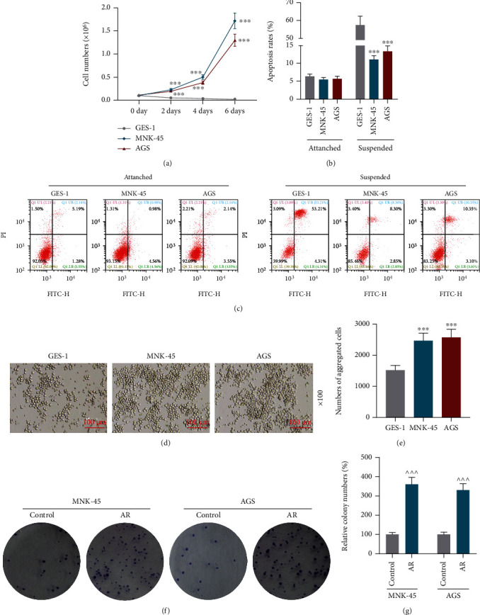
Figure 1.

Gastric cancer cells have stronger anoikis resistance compared with normal gastric mucosa epithelial cells. (a) Numbers of GES-1, MNK-45 and AGS cells were counted by a hemacytometer after suspension culture for 0, 2, 4, and 6 days. (b) Apoptosis rates of GES-1, MNK-45 and AGS cells after 24-hour adherent culture or suspension culture were quantitated through cell apoptosis assay. (c) Cell apoptosis was evaluated using a flow cytometer and an Annexin-V-FITC Apoptosis Detection Kit with PI. (d) Pictures of the morphology of GES-1, MNK-45 and AGS cells in suspension culture were taken under a microscope. (e) Numbers of aggregated GES-1, MNK-45 and AGS cells in suspension culture were counted by microscopy. (f) Representative pictures of colonies in colony formation assay. (g) Relative cell colony numbers were counted manually. ∗p <0.05 vs. GES-1 cells; ∗∗∗p <0.001 vs. GES-1 cells and ^^^ p <0.001 vs. control group. Data were performed as the means ± standard.
