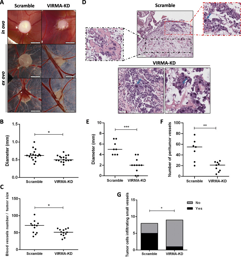
Fig. 5.
Knockdown of VIRMA attenuates the malignant phenotype in vivo. A – Macroscopic view of tumor formation (in ovo and ex ovo) and neo-angiogenesis in NCCIT scramble and VIRMA knockdown experimental conditions. Notice reduced size and decreased number of vessels in VIRMA knockdown tumors; B and C – Distribution of macroscopic tumor size and number of peri-tumor vessels (normalized to size of tumor) in scramble and VIRMA knockdown conditions; D – Histological aspects of scramble and VIRMA knockdown tumors. Notice the highly vascularized tissue around the scramble tumor, and infiltration of individual clusters of tumor cells around and into blood vessels (black box inset). Notice the cellular atypia (red box inset), including mitotic figure (blue arrow), in a more cellular area. The two VIRMA knockdown tumors with higher tumor cellularity are also shown (notice the clear cellular atypia, with prominent nucleoli and mitotic figures – blue arrows – including an atypical tripolar mitosis); E and F - Distribution of tumor size and number of peri-tumor vessels assessed histologically in scramble and VIRMA knockdown conditions; G – Presence of images of invasion into blood vessels. * p < 0.05; ** p < 0.01; *** p < 0.001

