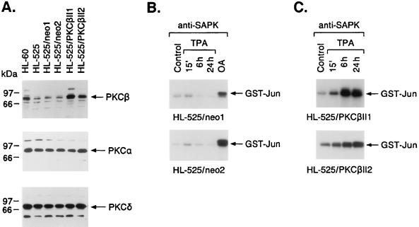
FIG. 6.
TPA-induced activation of SAPK in HL-525 cells stably expressing PKCβII. (A) HL-525 cells were stably transfected with pEF2/neo or pEF2/PKCβII. After selection, lysates were subjected to immunoblotting with anti-PKCβII, anti-PKCα, and anti-PKCδ antibodies. (B and C) HL-525/neo (B) and HL-525/PKCβII (C) clones were treated with 16 nM TPA for the indicated times. Anti-SAPK antibody immunoprecipitates were assayed for GST-Jun phosphorylation. Cells were also treated with 40 ng of OA per ml for 6 h.

