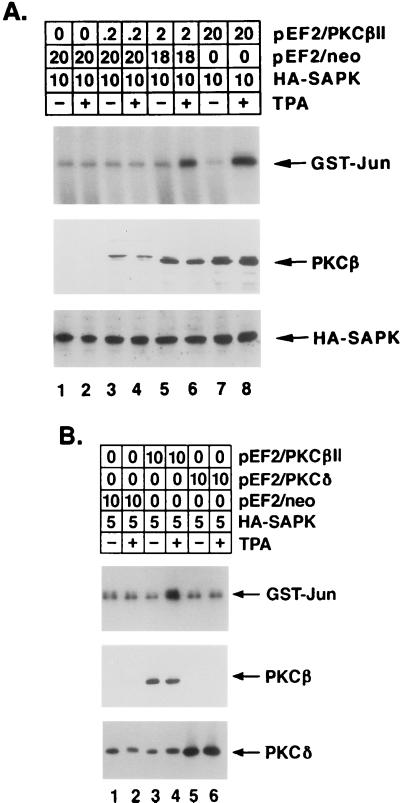
FIG. 9.
PKCβII-dependent SAPK activation in TPA-treated HeLa cells. HeLa cells were transfected with the indicated amounts (micrograms) of pEF2/PKCβII, pEF2/PKCδ, pEF2/neo, and HA-tagged SAPK. At 48 h posttransfection, the cells were left untreated or were treated with 16 nM TPA for 15 min. Cell lysates were immunoprecipitated with anti-HA antibody, and the anti-HA antibody immunoprecipitates were assayed for phosphorylation of GST-Jun. Lysates were also subjected to immunoblot analysis with anti-PKCβII, anti-HA, and anti-PKCδ antibodies to assess the levels of expression of transfected PKCβII, HA-tagged SAPK, and PKCδ (lower panels). Panel A shows a dose dependence on PKCβII expression level, and panel B shows the specificity of PKCβII in comparison with PKCδ.

