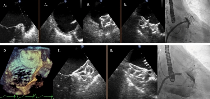
Fig. 1.
a1 and a2 Transesophageal echocardiography (TEE) of the left atrial appendage in X-plane view (65/-24). b1 and b2: the image shows a non-appropriate orientation and apposition with the 28 mm AMULET device. c Angiography showing a lack of compression on the device and although there was no contrast inside it was decided not to release the device. With the use of FEops application a 34 mm Amulet device was implanted and released after checking good apposition and compression. d tridimensional TEE image of the device. e1, e2 and f TEE and angiographic images showing correct colocation of the Amulet device

