Fig. 2.
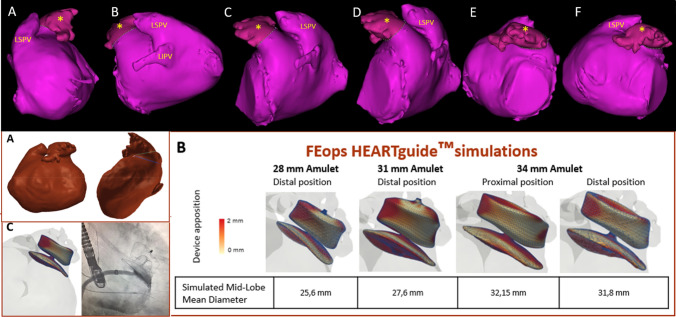
Top row: Superior CT reconstruction of the left atrial appendage (LAA) (*), seen progressively rotating from extreme posterior (a–f) to anterior projection counterclockwise. LSPV-left superior pulmonary vein. LIPV-left inferior pulmonary vein. Bottom row. Analysis received from FEops application. a Left atrial and LAA seen in anterior and lateral LAA projections. Blue lines: ostium and landing zone measurements of LAA. b Simulations with 3 sizes of Amplatzer™ Amulet™ devices 28, 31 and 34 mm. From this last one, proximal or distal implant and apposition degree are also simulated. c Size (34 mm) and shape of the chosen implant (distal) and the result of the real implant in the procedure

