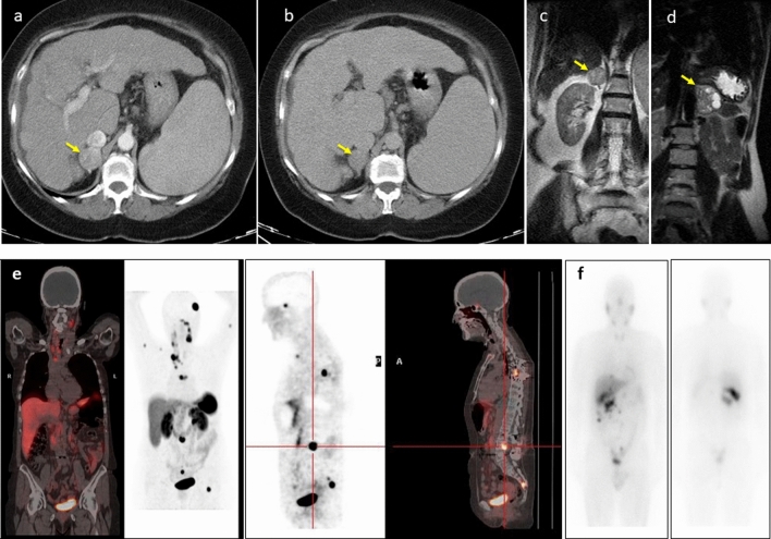
Fig. 2.
Typical morphological and functional imaging of PPGLs. a, b Axial contrast-enhanced CT portal (a) and delayed phase (b) of the upper abdomen showing the pheochromocytoma in the right adrenal gland (yellow arrowhead). Intravenous contrast administration typically enhances avidly due to the capillary-rich framework of the tumor. c, d Coronal T2-weighted MRI images revealed a homogeneous pheochromocytoma (c) in the right adrenal gland (yellow arrowhead), and other pheochromocytomas in the left adrenal gland (yellow arrowhead) with central necrosis are characteristically “light-bulb” bright lesions on T2-weighted imaging (d). Pheochromocytomas are potentially malignant (10%), and the only reliable criterion for the diagnosis of malignancy is metastatic spread. e A 61-year-old woman with metastatic cervical paraganglioma. 68 Ga-DOTATOC PET/CT study showing bilateral laterocervical lymph nodes, mediastinal involvement and multiple bone metastases. f A 56-year-old man was diagnosed with a 44 × 39-mm right adrenal incidentaloma. After right adrenalectomy, a histological study showed pheochromocytoma without evidence of malignancy. Negative genetic study. During follow-up, he presented with recurrence. Body scan with 123-I-MIBG shows lesions in the right renal cell and multiple peritoneal implants, some in contact with the liver surface without being able to rule out secondary infiltration. The patient has received treatment with 131I-MIBG with stabilization of the disease

