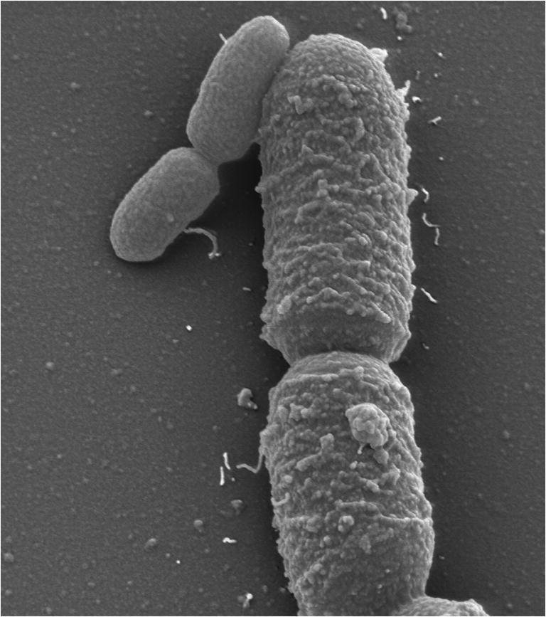
Fig. 1.

Electron microscope image of Priestia megaterium (large cells) and Escherichia coli (small cells). P. megaterium and E. coli were individually grown aerobically in rich medium at 37 °C, mixed in the middle of their exponential growth phases and examined in a field emission scanning electron microscope (FESEM) Zeiss DSM982 Gemini (magnification 6,500-fold). The picture was taken by Manfred Rohde, Helmholtz Centre for Infection Research, Braunschweig, Germany.
