
FIGURE 2.
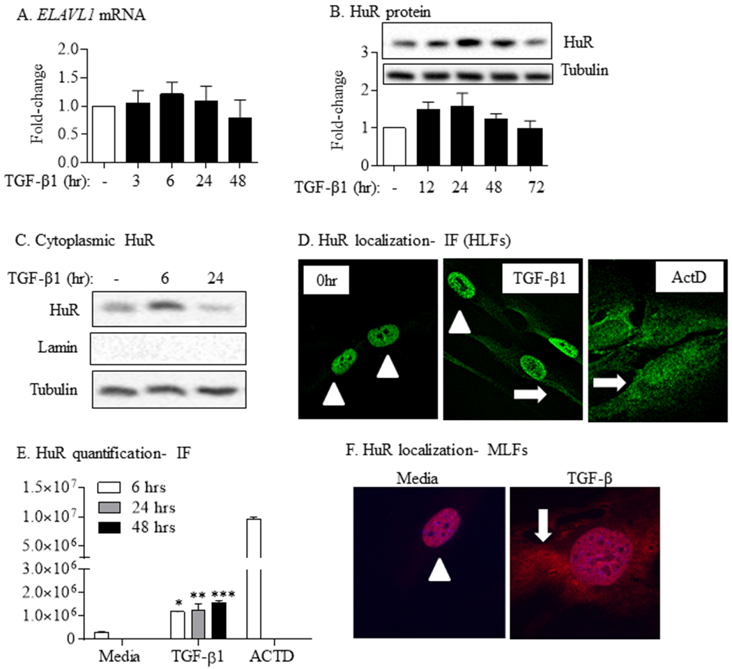
TGF-β1 increases cytoplasmic HuR localization in primary lung fibroblasts. TGF-β1 did not alter total HuR mRNA (ELAVL1) (A) or protein (B) expression. Representative western blot is shown. Note the same tubulin is also presented in Figure 1A and 1B as the HuR western blot selected was from the same experiment/western blot as that selected for α-SMA and COL1A1. Values are means ± SEM (n = 6). (C) Cytoplasmic HuR was increases in response to TGF-β compared to media-only cells. Tubulin was used as a housekeeping and Lamin for cytoplasmic purity. (D) HuR localization- IF (HLFs): HuR is predominantly nuclear (arrowheads) in media-only cells (0 hr). In response to TGF-β1, there is robust translocation of HuR to the cytoplasm (arrow). ActD was used as a positive control; note the re-distribution to the cytoplasm in response to ActD (arrows). (E) HuR quantification-IF: there was a significant increase in HuR cytoplasmic levels in response to TGF-β at all time-points. (F) HuR localization- MLFs: there was also translocation of HuR to the cytoplasm (arrow) in primary MLFs treated with TGF-β. Representative images are shown.