
FIGURE 6.
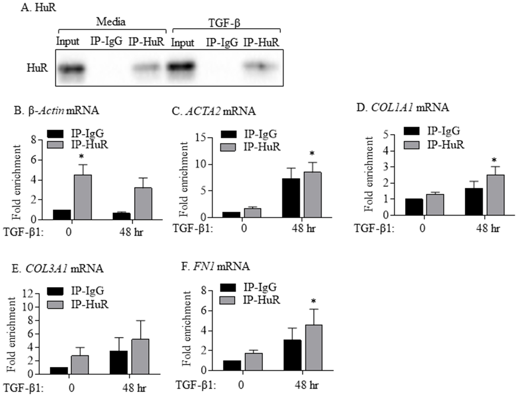
Binding of HuR to mRNAs of ACTA2 and ECM genes in HLFs treated with TGF-β1. (A) HuR: Representative western blot of HuR IP in HLFs treated with TGF-β1 for 48 hrs. Input refers to crude cell lysates. IP-IgG refers to immunoprecipitation (IP) with control IgG antibody while IP-HuR refers to the IP with anti-HuR IgG antibody. Note the presence of HuR protein in IP-HuR but not in IP-IgG. Detection of mRNA for β-Actin (B; positive control), ACTA2 (C), COL1A1 (D), COL3A1 (E), and FN1 (F) in IP-IgG and IP-HuR was done using qPCR. Values are expressed as fold change to values measured in IP-IgG in HLFs untreated with TGF-β1 (0 hr). Note the enrichment of β-Actin, ACTA2, COL1A1, and FN in IP-HuR of cells treated with TGF-β1. Results are presented as the mean ± SEM (n = 3 independent experiments) (p< 0.05 compared to IP-IgG).