
FIGURE 9.
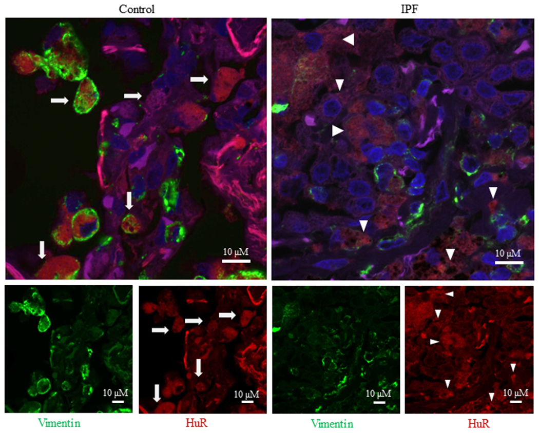
Cytoplasmic HuR in IPF lung. There is extensive cytoplasmic localization of HuR in IPF lung. A representative slide is shown of control (left panel) and IPF (right panel) lung that was stained for vimentin (green), HuR (red) or DAPI (blue); the larger merge image is shown above. Note the extensive cytoplasmic localization of HuR to fibroblasts in the IPF lung (arrowhead) compared to the largely nuclear HuR (arrows) in the control (non-fibrotic) lung.