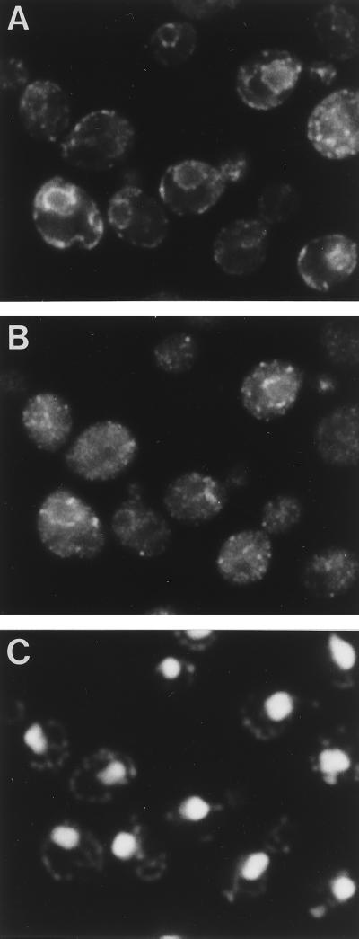
FIG. 11.
Double immunofluorescence staining of BiP and 3HA-Rer2p. Δrer2 cells (SMY41) harboring 3HA-RER2 on a single-copy plasmid were grown at 30°C and prepared for indirect immunofluorescence microscopy. (A) Rhodamine fluorescence corresponding to the anti-BiP antibody. (B) Fluorescein fluorescence corresponding to the anti-HA antibody (16B12). (C) DNA staining with DAPI.

