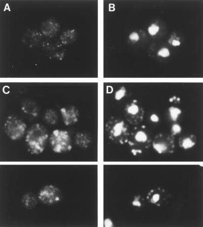
FIG. 4.
Immunofluorescence localization of a cis-Golgi marker protein, Ypt1p. Wild-type (SNY9) (A and B) and rer2-2 (SNH23-7D) (C and D) cells were incubated at 37°C for 4 h and subjected to indirect immunofluorescence microscopy with the anti-Ypt1p antibody. (A and C) Fluorescence images with the antibody. (B and D) DNA staining with DAPI.

