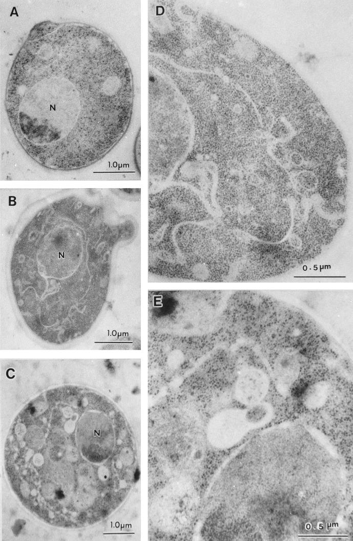
FIG. 5.
Electron micrographs of rer2 cells. Wild-type (SNY9) (A) and rer2-2 (SNH23-10D) (B to E) cells were incubated at 23°C (A, B, and D) or shifted to 37°C for 2 h (C and E) and then subjected to freeze-substitution fixation and electron microscopic observation. Panels D and E are enlargements of panels B and C, respectively.

