FIG. 9.
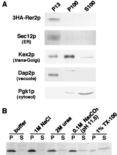
(A) Subcellular fractionation of 3HA-Rer2p. Δrer2 cells expressing 3HA-Rer2p on a single-copy vector were spheroplasted, homogenized, and subjected to a series of centrifugations: 300 × g for 5 min, 13,000 × g for 15 min, and 100,000 × g for 45 min. Aliquots were taken from the pellet of the 13,000 × g centrifugation (P13) and the pellet (P100) and supernatant (S100) fractions of the 100,000 × g centrifugation and analyzed by immunoblotting. (B) Extraction of 3HA-Rer2p. The total homogenate was treated with the reagents indicated and centrifuged at 436,000 × g for 1 h. The pellets and supernatants were analyzed by Western blotting with the anti-HA antibody. TX-100, Triton X-100.

