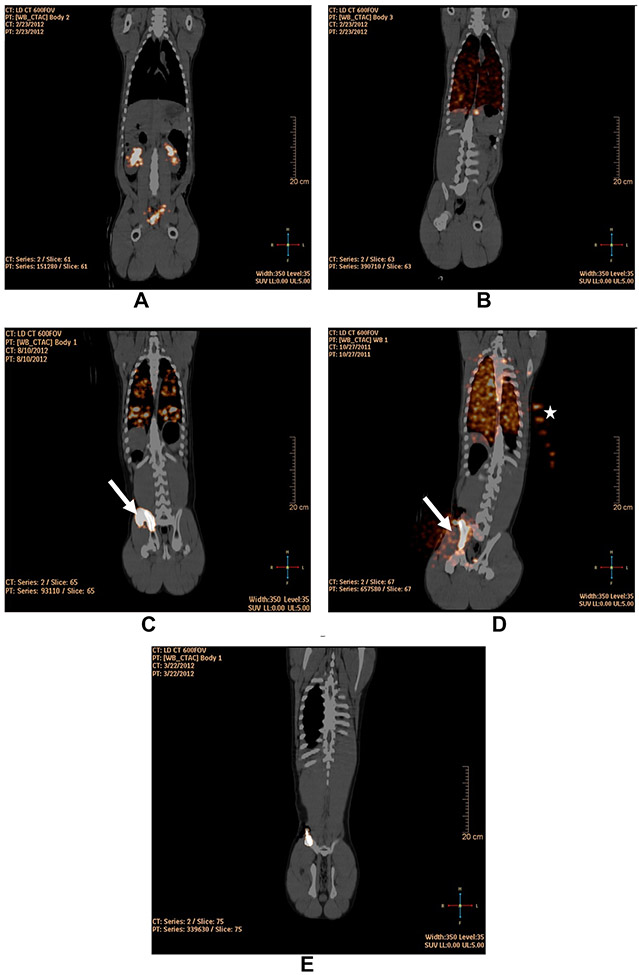
Figure 4:
Fusion PET/CT coronal images at 1 h following (A) IB-injection of 89Zr-Df with radioactivity in the renal pelvises and bladder (Pig ID# 32890); (B) IV-injection of 89Zr-hCD133+ with radioactivity confined to the lung (Pig ID#32891); (C) Hand IB-injection of 89Zr-hCD133+ into two sites on the right iliac-crest demonstrating radioactivity leaked at the first site of injection (arrow) and radioactivity within the iliac-BM and lungs (Pig ID#34489); (D) hand IB-injection of 89Zr-hCD133+ into a single site on the right iliac-crest demonstrating radioactivity leaked at the sight of injection (arrow) and radioactivity within the iliac-BM and lungs (Pig ID# 31717); (E) slow non-manual IB-infusion (0.2 mL/min) of 89Zr-hCD133+ into the right iliac-crest demonstrating radioactivity confined within the iliac-BM (Pig ID# 33189). Note that in (D) there is radioactivity highlighted in syringes of 89Zr-oxalate placed to the left of the swine for calibration and decay correction (star).

