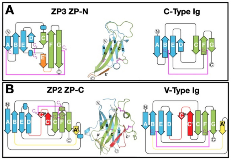
Figure 4.
Three-dimensional structures of ZPD subdomains ZP-N and ZP-C that are related to C- and V-type Ig-like domains. (A) ZP3 subdomain ZP-N and C-type Ig-like domains. β-strands are labeled using Ig terminology; helices are indicated by rectangles. Opposing β-sheets 1 and 2 are blue and green, respectively, with termini circled. The E′ strand is orange and disulfides magenta. (B) ZP2 ZP-C and V-type Ig-like domains. As in panel A, except for the additional A′ and C′/C″ strands that are yellow and red, respectively. This figure was adapted with permission from L. Jovine ([11], Figure 4), Copyright 2018.

