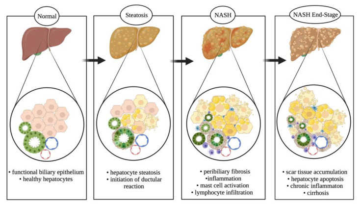
Figure 4.
NAFLD spectrum. NAFLD progresses from a healthy liver parenchyma (steatosis < 5% of hepatocytes) to a stage (steatosis > 5% of hepatocytes) with initiation of ductular reaction, mast cells and macrophage infiltration, and peribiliary fibrosis coupled with biliary senescence. It progresses further to a severe stage of NASH with scar tissue accumulation, elevated steatosis and hepatic ballooning, and increased circulating cytokine and SASP secretion from senescent bile ducts. Upon persistent dietary conditions, it assumes a chronic phenotype resulting in hepatocellular carcinoma and cirrhosis. Image made with BioRender (https://biorender.com/).

