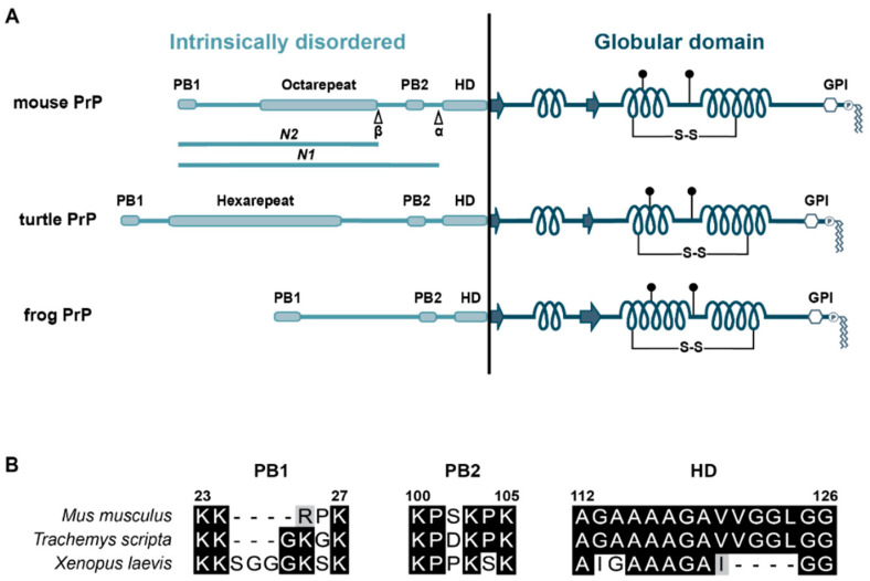
Figure 1.
(A) Schematic representation of PrPC from mouse, turtle, and frog. PB: polybasic motif; β: cleavage site that generates N2; α: cleavage site that generates N1; HD: hydrophobic domain; arrows: β-strand; coils: α-helices; S-S: disulfide bridge; filled circles: N-linked glycans; GPI: glycosylphosphatidylinositol anchor. (B) Sequence alignments of PB1, PB2, and the HD. Identical residues are marked in black, similar residues in gray (GenBank accession numbers: M18070.1, XP_034617687.1, AAH94089.1). The numbering of the residues refers to mouse PrPC.

