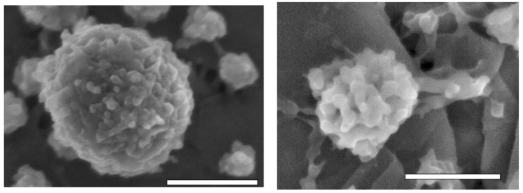
Figure 4.
SEM micrographs of intact (left) and dissociated (right) casein micelles. Scale: 200 and 100 nm, respectively [49]. Reproduced with permission from Douglas G. Dalgleish, Paul A. Spagnuolo, H. Douglas Goff, A possible structure of the casein micelle based on high-resolution field-emission scanning electronmicroscopy; published by Elsevier, 2004.

