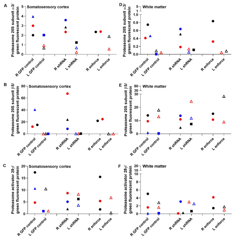
Figure 11.
Western blot data for proteasome 20S subunits in piglets that received intracerebral adeno-associated virus (AAV)-green fluorescent protein (GFP), AAV-shRNA to PA28γ-GFP, or AAV-PA28γ-GFP and sham procedure with hypothermia. Data are normalized to (divided by) the piglet’s GFP level as a measure of AAV transduction in the right (R) and left (L) somatosensory cortex (A–C) and subcortical white matter (D–F). AAV-shRNA to PA28γ-GFP did not consistently decrease PA28γ levels or alter the levels of α3 and β5 subunits compared to AAV-GFP. AAV-PA28γ-GFP did not reliably raise the subunit levels. Data points from the same piglet are matched by color within each treatment group. The shapes indicate the AAV dose in each cerebral hemisphere: square = 2 × 10¹⁰–2 × 10¹¹ genome copies (gc), circle = 4 × 10¹⁰–4 × 10¹¹ gc, and triangle = 5 × 10¹⁰–5 × 10¹¹ gc. Open symbols indicate piglets that also received adjuvant (40 ng of F108 and 2.5 µg of polybrene). Data were divided by a Ponceau-stained protein loading control.

