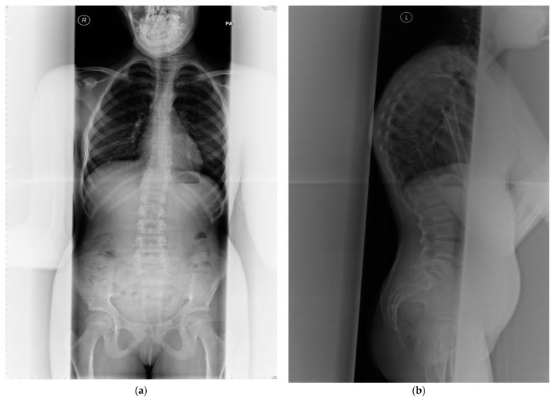
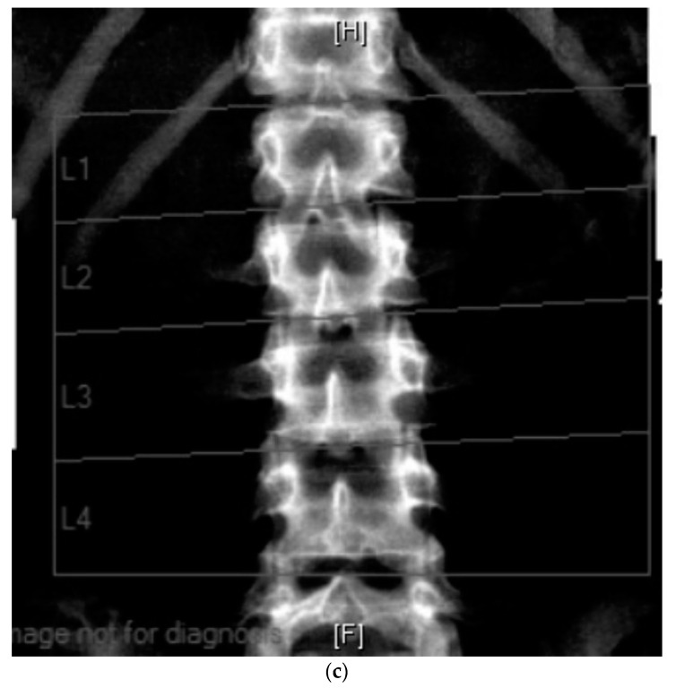
Figure 2.
(a) Anterior posterior view—whole spine x-ray in a 25-year-old female patient affected with PMM2-CDG; bone mineral density is below expected for her age (T score −2.6, z-score −2.6); (b) lateral view; (c) radiological features of her lumbar spine: there is a double mild curve scoliosis and no rotational deformity. All pedicles are visualized. There is impression of slight upper hyperkyphosis and impression of multilevel Schmorl’s nodes.


