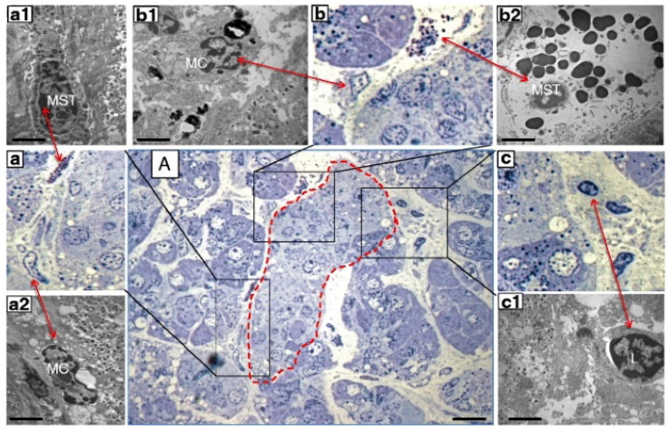
Figure 2.
Consecutive semithin and ultrathin sections of a pancreas sample from a donor with type 1 diabetes showing (A) a pancreatic islet (dashed line) surrounded by infiltrate containing different inflammatory cells (×1000). The comparison between consecutive semithin and ultrathin sections illustrates how the identification of the different types of inflammatory cells in the semithin section was confirmed by electron microscopy. (a–c) ×1000 magnification (semithin sections) of the corresponding images in (A); (a1,a2) electron microscopy images of a mast cell (MST) and macrophage (MC) identified in (a) (×10,000); (b1,b2) electron microscopy images of an MC and degranulating MST identified in (b) (×10,000); (c1) electron microscopy of a lymphocyte (L) identified in (c) (×10,000). Scale bars correspond to 10 μm in A, and to 1 μm in (a1,a2,b1,b2,c1). Reproduced with permission from Ref. [62].

