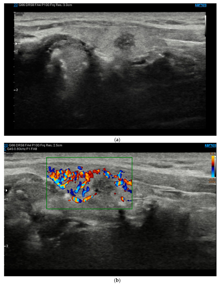
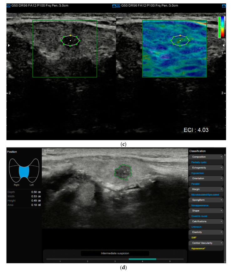
Figure 1.
(a). At B-mode Ultrasound (US), the lesion appeared round-shaped, hypoechoic with irregular margins (EU-TIRADS 5). (b). At color–Doppler US evaluation, the lesion showed no internal or peripheral vascularization (pattern I). (c). At US-Elastography (USE) evaluation, the lesion appeared stiff (ECI: 4.03). (d). At S-Detect evaluation, the lesion suggested intermediate suspicion of malignancy (TIRADS 4). Finally at histology it was identified as a papillary carcinoma.


