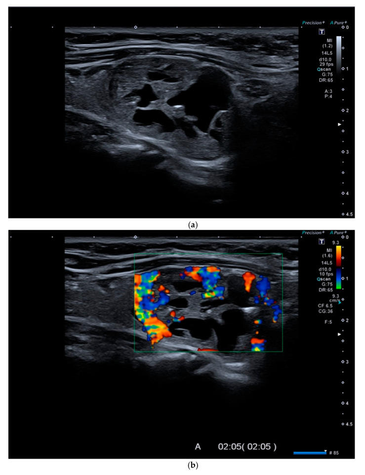
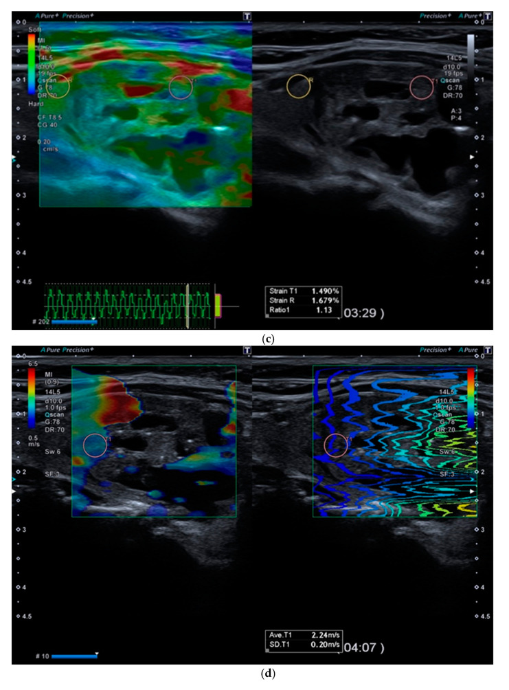
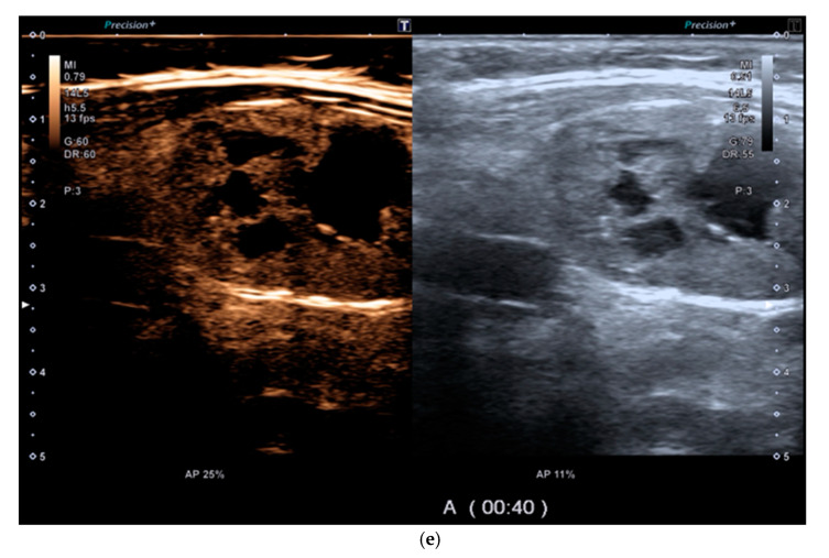
Figure 2.
(a). At B-mode Ultrasound (US), an oval-shaped nodule with mixed ecostructure, some internal fluid areas and smooth margins, was identified (EU-TIRADS 3). (b). At color–Doppler US evaluation, the lesion appeared with internal and peripheral vascularization (pattern III). (c). At Strain Ratio Elastography (SRE) evaluation, the lesion appeared soft (SR 1.13). (d). At Shear Wave Elastography (SWE) evaluation, the lesion appeared soft (2.24 m/s). (e). At CEUS evaluation, the lesion appeared solid and richly vascularized, similar to the surrounding thyroid parenchyma without wash-out. At histology, the lesion was confirmed to be a follicular hyperplasia.


