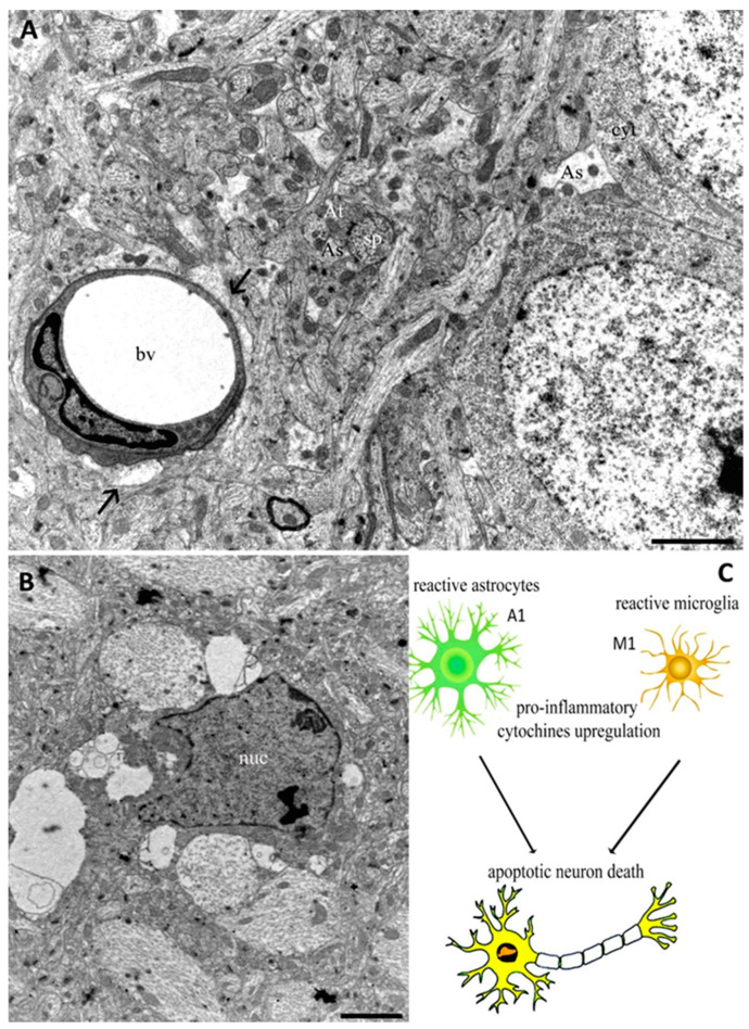
Figure 1.
Transmission Electron Microscopy (TEM) images from cerebral cortex of adult rat (A,B). In (A), astrocyte end feet protrusions (arrows) can be observed near to a blood vessel (bv). Astrocyte processes (As) appear near axonal terminal (At) and dendritic spines (sp), as well as around neuron cytoplasm (cyt). The micrograph (B) shows a microglia cell characterized by a nucleus (nuc) with an irregular outline in which the chromatin is clumped beneath the nuclear envelope. The schematic representation of glia activation related to neuron death appears in (C). Bars 2 µm for (A) and (B).

