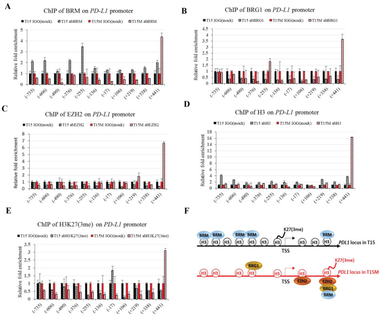
Figure 6.
The chromatin status of the PD-L1 locus varies upon CD4+ T cell exhaustion: (A) the presence of BRM ATPase of SWI/SNF CRCs on the PD-L1 locus; (B) the presence of BRG1 ATPase of SWI/SNF CRCs on the PD-L1 locus; (C) the presence of the EZH2 subunit of PRC2 on the PD-L1 locus; (D) the presence of histone 3 (H3) on the PD-L1 locus; (E) the presence of histone 3 lysine 27 trimethylation (H3K27me3) on the PD-L1 locus; (F) model describing chromatin status on the PD-L1 locus during exhaustion of CD4+ T cells. T15; CD4+ T cells restimulated for three days; T15M; CD4+ T cells restimulated in co-culture with cancer cells for 3 days. The experiment was repeated 3 times.

