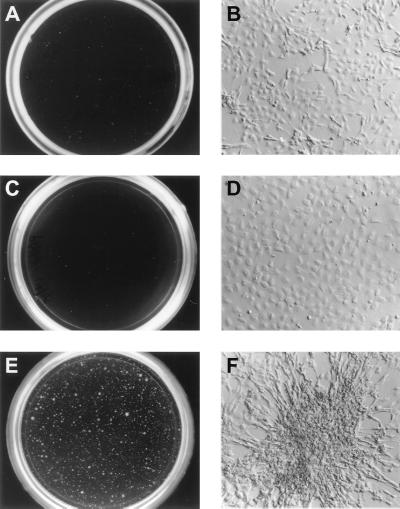
FIG. 4.
Cell morphology of NIH 3T3 cells infected with high-titer retroviral stocks containing pSRα(ΔHindIII)-tk-Neo (A and B), Pax3 (C and D), or Pax3-FKHR (E and F). Cells were grown on plastic, and colonies were visually scored 2 and 3 weeks after plating. Colonies shown are representative of the total population.

