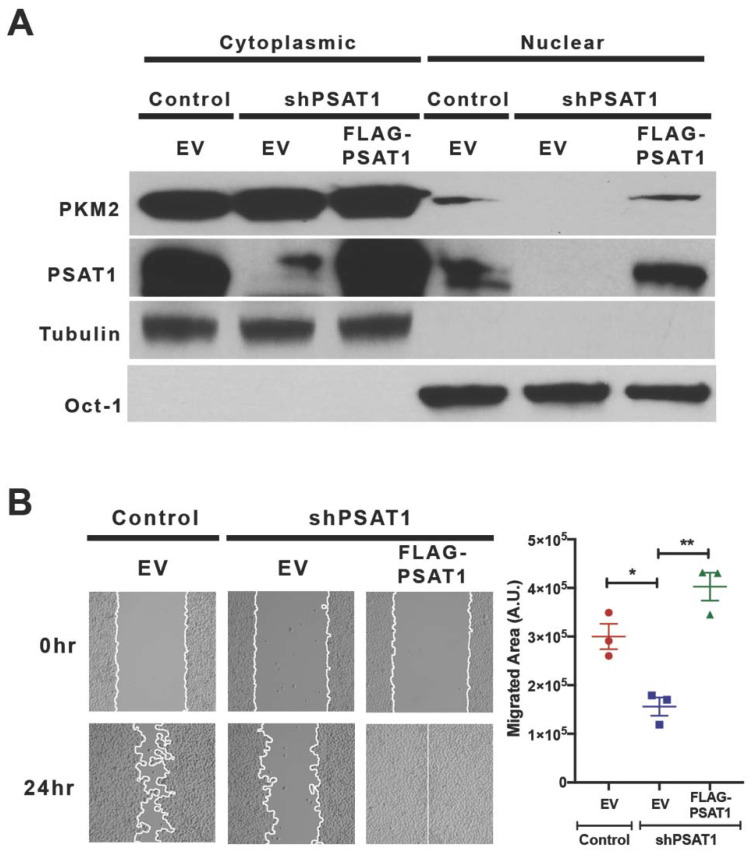
Figure 6.
Re-expression of PSAT1 restores the nuclear localization of PKM2 and cell migration in silenced PC9 cells. (A) Immunoblot analysis for PKM2 and PSAT1 localization in PSAT1-silenced PC9 cells stably expressing an empty vector (EV) or FLAG-PSAT1. Cytoplasmic and nuclear fractions from the control-EV, shPSAT1-EV, and shPSAT1-FLAG-PSAT1 PC9 cells were analyzed using anti-PKM2 and anti-PSAT1 antibodies. Oct-1 and α-tubulin served as loading controls for the nuclear and cytoplasmic compartments, respectively. Shown are representative images from three independent experiments. (B) Wound healing assay of the control-EV, shPSAT1-EV, and shPSAT1-FLAG-PSAT1 PC9 cells. Shown are representative images at 0 h and 24 h. The migrating cells are demarcated by continuous white lines. Data are presented as a mean ± SE migrated area after 24 h from three independent experiments. A statistical significance was determined by a one-way ANOVA with Tukey’s multiple comparison test. ** p < 0.005 and * p < 0.05. A.U.: arbitrary unit.

