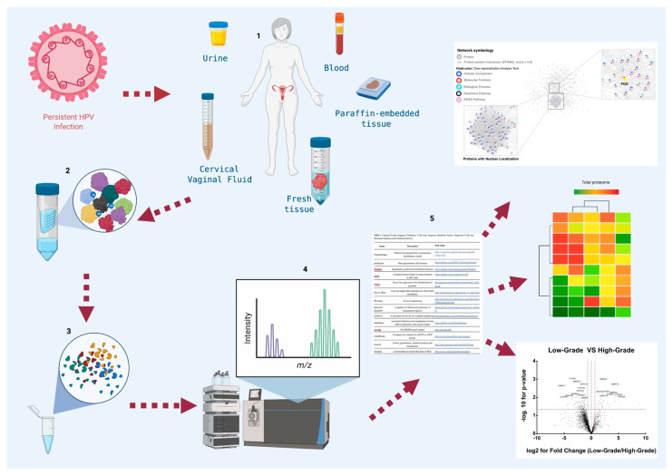
Figure 1.
General diagram of the workflow in proteomic studies in cervical cancer. Proteomic studies in cervical cancer are designed to search for biomarkers that could be used in the diagnosis, prognosis, and identification of new therapeutic targets, and they are carried out Figure 1. (1) Collection of biological samples from patients and controls such as: urine, vaginal cervical fluid, blood, and tumor tissue. If the samples start from cells, it is essential to perform cell lysis or mechanical destruction (freeze thaw, French press, sonication macerated with liquid N2, lysis with detergents). (2) Purification of total proteins by centrifugation. The storage of the sample until its use at temperatures of −20 °C or −70 °C with or without protease inhibitor according to the chosen step of proteomic analysis to avoid any interference with the selected method. (3) Proteomic strategy selection, based on advanced techniques: protein microarray, mass spectrometry, Edman sequencing, 2D gel, 2D-DIGE. Quantitative techniques: ICAT, SILAC, iTRAQ. High throughput techniques: X-ray crystallography, NMR spectroscopy. (4) Match in databases and validation of candidates by ELISA, Western Blot or Immunohistochemistry. (5) Graphics construction. Partially created with BioRender.com.

