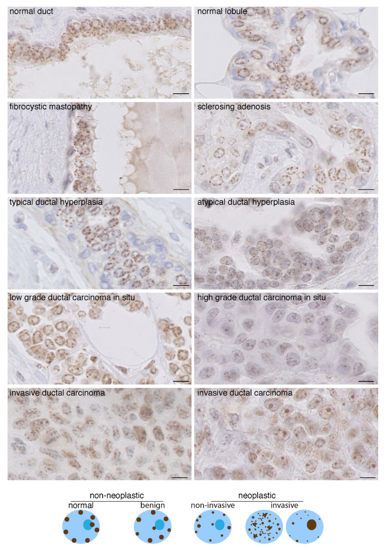
Figure 3.
CENP-A localization patterns in breast lesions. Top: CENP-A staining by IHC in breast tissues as indicated. Invasive ductal carcinoma displaying CENP-A foci localized only inside the nuclear space (left) and a few remaining at the nuclear periphery (right) are shown. Scale bar is 10 µm. Bottom: scheme depicting the patterns.

