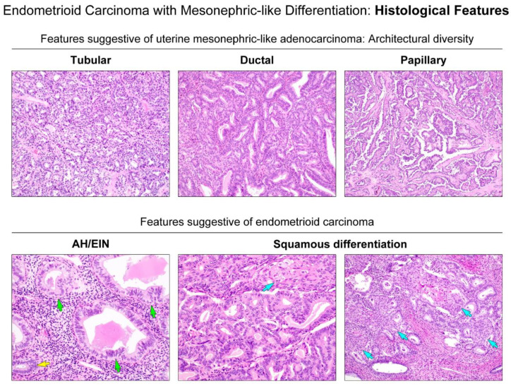
Figure 3.
Histological features suggestive of either uterine MLA or endometrial EC. (Upper panels) Diverse growth patterns, including tubular, ductal, and papillary architecture, are suggestive of uterine MLA. (Lower panel) The presence of atypical hyperplasia/endometrioid intraepithelial neoplasia (AH/EIN) and foci of squamous differentiation (blue arrows) are suggestive of endometrial EC. Compared to the nuclei of non-atypical glands (yellow arrows), AH/EIN (green arrows) shows a definitive cytological demarcation, including nuclear pleomorphism, enlargement, rounding, and loss of polarity.

