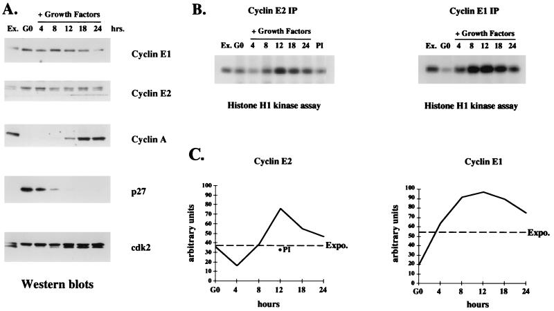
FIG. 6.
Cyclin E2-associated kinase activity is cell cycle regulated. (A) Western blot analyses with cell extracts harvested at the times indicated following growth factor stimulation of quiescent human MCF10 cells. Ex., exponentially proliferating cells; G0, quiescent cells at time zero. The membranes were probed with antibodies for cyclin E1, cyclin E2, cyclin A, p27Kip1, and Cdk2 expression. (B) Histone H1 kinase assays performed with cyclin E1 and cyclin E2 immunoprecipitates (IP) from the same extracts as used in the Western blots depicted in panel A. PI indicates kinase activity present in preimmune serum immunoprecipitates from MCF10 extracts harvested 12 h after growth factor addition. (C) Relative intensities of phosphorylated histone H1 bands determined by densitometric scanning. The intensity of each band is depicted on the y axis in arbitrary units, 0 indicates quiescent cells, and Expo. indicates the cyclin-associated kinase activity in exponentially growing cells. For the cyclin E2 analysis, the level of kinase activity in preimmune immunoprecipitates at 12 h is depicted with a filled circle.

