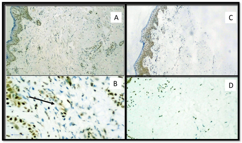
Figure 4.
(A) Immunostaining for HMGB1 of a patient positive for SARS-CoV-2: note the internal positive control of normal epidermal keratinocytes and positivity in the cells constituting the excretory portion of the eccrine sweat glands that were strongly positive for anti-S1 spike protein immunostaining by SARS-CoV-2 (Immunohistochemistry, Original Magnification: 40×). (B) Detail of extracellular release of HMGB1 (Immunohistochemistry, Original Magnification: 200×). (C) Immunostaining for HMGB1 of a patient negative for SARS-CoV-2: note the internal positive control and substantial negativity in the cells of superficial dermis (Immunohistochemistry, Original Magnification: 40×). (D) Detail of negativity for HMGB1 (Immunohistochemitry, Original Magnification: 100×).

