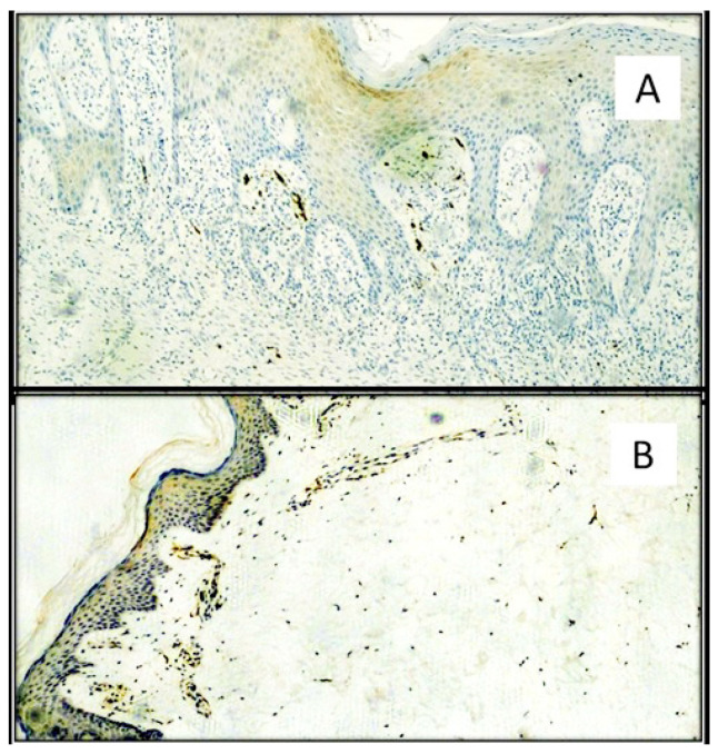
Figure 8.
(A) Immunostaining for HO-1: note the positivity of a few inflammatory cells available mainly subepithelial in the skin of a patient positive for SARS-CoV-2 (Immunohistochemistry, Original Magnification: 40×). The light brown color is linked to “background noise” of the immunohistochemical reaction. (B) Photomicroghaph of skin biopsy of a patient negative for SARS-CoV-2 with higher expression of HO-1 in the epidermis and of inflammatory cells in subepithelial disposition (Immunohistochemistry, Original Magnification: 100×).

