Figure 7.

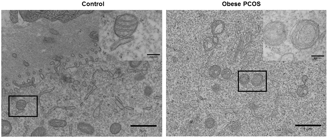
Transmission Electron Microscopy of Oocytes: Transmission electron microscopy image of control showing normal mitochondrial structure (A) and obese PCOS showing compromised structure with abnormally formed cristae (B). n=4/group.

Transmission Electron Microscopy of Oocytes: Transmission electron microscopy image of control showing normal mitochondrial structure (A) and obese PCOS showing compromised structure with abnormally formed cristae (B). n=4/group.