Figure 3.
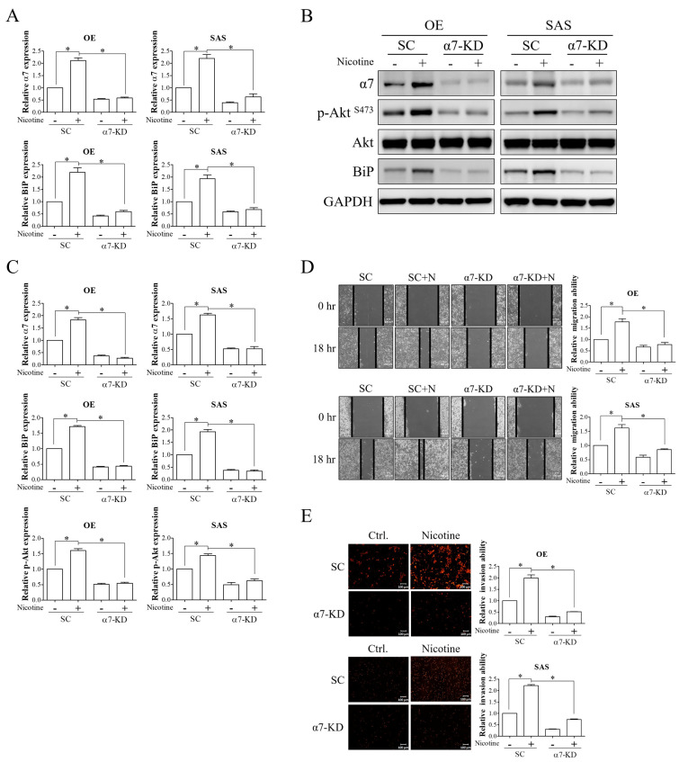
α7-nAChR-Akt signaling was involved in nicotine-induced BiP expression and malignant behaviors in OSCC. OE and SAS cells with/without α7-nAChR silencing were treated with 1 μM nicotine for 48 h. (A,B) The expressions of α7-nAChR and BiP and activation of Akt, as indicated by the expression level of phospho-Akt at Ser473, were analyzed by quantitative RT-PCR and Western blot analysis. (C) The graphs show the quantification of Western blots. Band intensities were quantified using ImageJ software. The relative protein expressions of α7-nAChR and BiP were normalized to GAPDH expression. The relative protein expression of phospho-Akt (Ser473) was normalized to total Akt expression. The migratory (D) and invasive (E) abilities were examined by wound healing and Transwell invasion assays. Representative images are shown, and black solid lines indicate the wound borders acquired at 0 and 18 h after scratching (D, left panels). Quantitative results of migratory cells (D, right panels) and PI-stained invasive cells (E, right panels) were determined using ImageJ software. The ability of migration was calculated by the area reduction at 18 h compared to the wound area at 0 h. N, nicotine. SC, non-targeting siRNA-transfected cells. α7-KD, α7-nAChR siRNA-transfected cells. GAPDH was used as the loading control. * p < 0.05 by one-way ANOVA followed by Bonferroni’s post hoc test. Data are presented as the mean ± SEM of three independent experiments. SEM, error bars. Scale bar, 100 μm.

