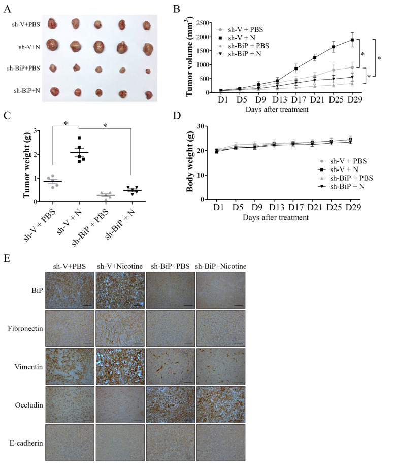
Figure 6.
BiP inhibition suppressed nicotine-induced oral cancer progression in nude mice. SAS cells transfected with scramble shRNA control (SAS-shV) or BiP shRNA (SAS-shBiP) (1.5 × 10⁶ cells/mice) were subcutaneously implanted into the right flank of nude mice. When the tumor size was about 100 mm3, the tumor-bearing mice were given daily intraperitoneal injections of either PBS or 1 mg/kg nicotine for one month. Following treatment, the mice were sacrificed and the tumor tissues were subjected to immunohistochemical staining for BiP and epithelial–mesenchymal transition (EMT) markers. (A) Representative images of dissected tumors (n = 5) from the nude mice. Average tumor growth curve (B) and tumor weight (C) in each group of mice (n = 5) were also recorded during treatment and at the time of mice sacrifice, respectively. The xenograft tumor volumes were measured twice every week using Vernier calipers and calculated using the formula: volume = (length × width2)/2. (D) The average body weight change of the nude mice. Bodyweight was measured twice every week. (E) Representative immunohistochemical staining images of the expressions of BiP, mesenchymal (fibronectin and vimentin), and epithelial (occludin and E-cadherin) markers in tumor tissues. N, nicotine. * p < 0.05 by one-way ANOVA followed by Bonferroni’s post hoc test. Data are presented as the mean ± SEM of 5 mice in each group. SEM, error bars. Scale bar, 100 μm.

