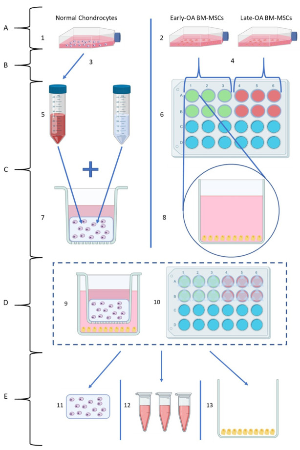
Figure 1.
Overview of the co-culture model (created using Biorender.com). (A) Recovery of cells from long-term storage: (1) Chondrocytes were recovered from liquid nitrogen storage and cultured in monolayer. (2) Donor-matched BM-MSC populations recovered from liquid nitrogen storage and cultured in monolayer. (B) Cell Collection: (3) Once 80% confluent, chondrocytes were collected by trypsinisation, and the required number for the model was separated from the suspension. (4) Once 80% confluent, BM-MSCs were collected by trypsinisation and counted, and the required number for the model was separated from the suspension. (C) Individual cell preparation for co-culture model: (5) Chondrocyte suspension was combined with 4% agarose solution to form a 1:1 mixture. (6) Donor-matched early OA and late-OA BM-MSCs seeded in 24-well plates (top view). (7) Agarose–chondrocyte mixture pipetted into 24-well plate transwell inserts. (8) BM-MSCs in 24-well plate (side-view) with osteogenic differentiation media added. (D) Co-culture setup: (9) Co-culture model (individual well, side view). (10) Co-culture model (full plate, top view). (E) Harvest of the co-culture model for analysis at endpoints (day 0, day 7, day 14 and day 21): (11) Chondrocyte–agarose scaffolds were removed for gene expression, histological and biochemical analyses. (12) Conditioned medium was removed for analysis of secreted protein abundance by ELISA. (13) BM-MSCs removed for gene expression and biochemical analyses (data not shown).

