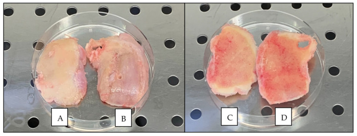
Figure 4.
Example of the macroscopic appearance of knee condyles collected from one patient following TKR surgery. (A) Condyle with the best (lowest) ICRS graded cartilage (cartilage side). (B) Condyle with the worst (highest) ICRS graded cartilage (cartilage side). (C) Condyle with the best ICRS graded cartilage (bone side). (D) Condyle with the worst (highest) ICRS graded cartilage (bone side). Note that cartilage is eroded down to the bone in the worst graded condyle (B).

