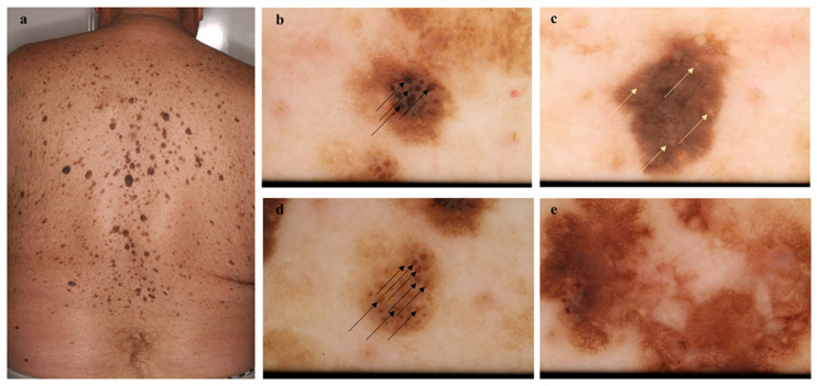
Figure 1.
Melanoma among several seborrheic keratoses (SK) on the back. This 67-year-old male patient was diagnosed with a melanoma on his back (in situ melanoma, pTis) near the shoulders and many SK lesions (a). Non-polarized dermoscopic images of SKs (b–d) and melanoma (e). SKs (b–d) show a dull surface, including fingerprint and cerebriform patterns, milia-like cyst (yellow arrows) and comedo-like opening (black arrows). Melanoma (e) contains an irregular pigment network and blue-white veil with multiple colors. The case of this patient also emphasizes the importance of full body examinations.

