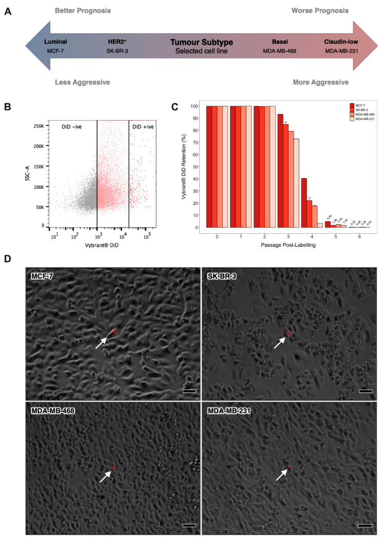
Figure 1.
Identification of Quiescent Breast Cancer Cells by Lipophilic Dye Retention In vitro. (A) Schematic depicting the prognosis of the main breast tumour subtypes and the representative cell line selected to model each of these. (B) Cytofluorimetric dot plot illustrating the gating strategy for identification of Vybrant® DiD retaining cells in adherent human breast cancer cell cultures after six passages of culture growth. Cytofluorimetric platforms were calibrated at the outset of each experiment using a Vybrant® DiD negative (DiD−) control sample and a Vybrant® DiD positive (DiD+) control sample that had been freshly labelled with Vybrant® DiD. (C) Proportion of label-retaining cells within adherent MCF-7, SK-BR-3, MDA-MB-468 and MDA-MB-231 cultures over six consecutive passages of culture growth as measured by flow cytometry. Data are expressed as mean ± SE (n = 3). (D) Phase-contrast and fluorescent image overlays of MCF-7, SK-BR-3, MDA-MB-468 and MDA-MB-231 cultures after 6 passages of culture growth post-staining with Vybrant® DiD (scale bar = 50μm). White arrows indicate DiD+ cells (red).

