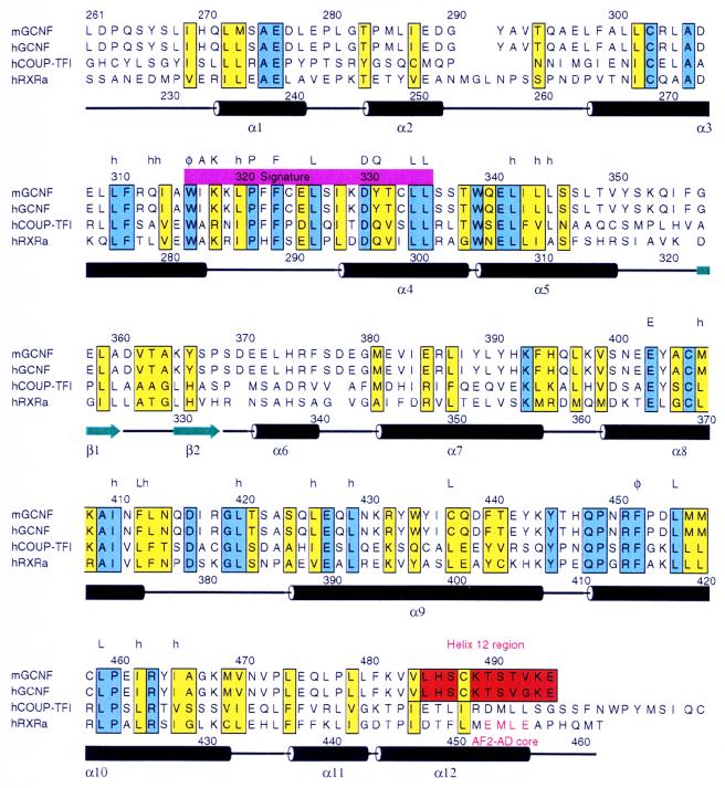
FIG. 1.
Alignment of the LBDs of mGCNF, hGCNF, hCOUP-TFI, and hRXRα. Conserved and similar residues are boxed in blue and yellow, respectively. The H12 region located at the very C terminus of the GCNF LBDs is colored in red. Secondary structure elements found in the crystal structure of the apo-hRXRα LBD (5) are indicated; α helices are depicted as black cylinders, and β strands are shown as green arrows. Regions of highest homology between mGCNF and hRXRα encompass H3 to H5 and H8 to H10. The position of AF2 AD core is indicated. Letters above the amino acid sequence of hRXRα mark residues that are highly conserved in the canonical fold of nuclear receptor LBDs (50). Abbreviations: h, hydrophobic; φ, aromatic; A, alanine; K, lysine; P, proline; F, phenylalanine; L, leucine; D, aspartic acid; Q, glutamine; E, glutamic acid.

