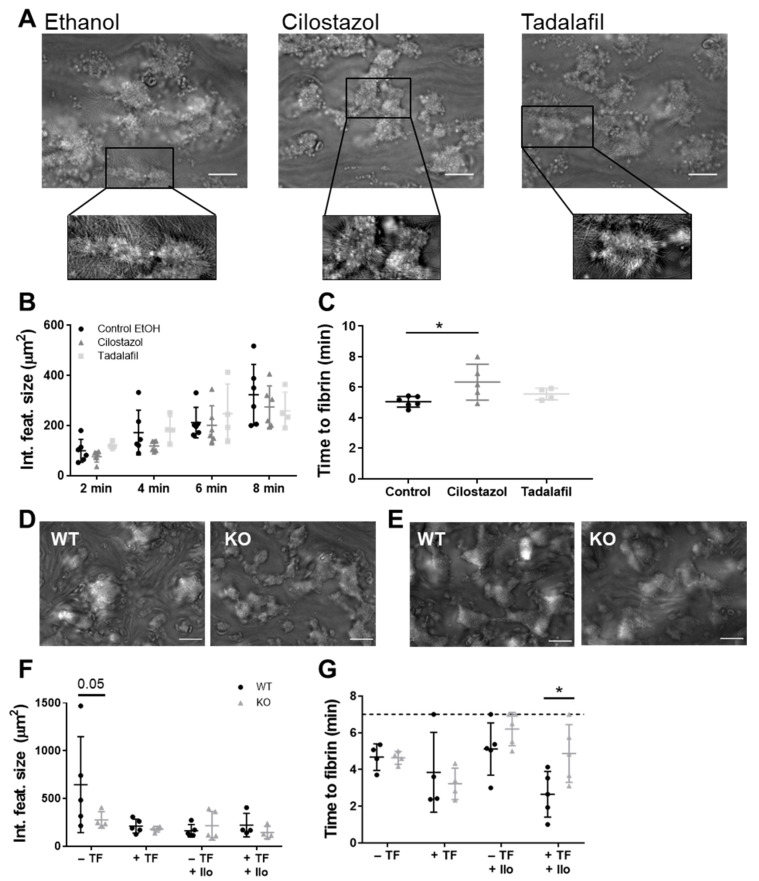
Figure 2.
Effects of PDE inhibition or genetic deletion on platelet-dependent coagulation under flow over collagen. Recalcified citrate-anticoagulated human (A–C) or mouse (D–G) blood was perfused over a collagen type I surface or a combined collagen type I plus tissue factor surface for 7 (mouse) or 8 (human) min at a wall shear rate of 1000 s−1. (A) Representative brightfield images after 8 min of blood perfusion without inhibitor (0.1% ethanol) or in the presence of cilostazol (50 μM) or tadalafil (100 nM). Quantitative analysis of integrated feature size (μm) (B) and time to fibrin formation (C). Representative brightfield images of blood perfusion of WT mice and Pde3a KO mice over collagen type I without (D) or with (E) tissue factor in the absence of iloprost under coagulating conditions. Quantitative analysis of integrated feature size (F) and time to fibrin (G). Scale is 20 μm. Mean ± S.D., n = 4–6 (human) or 4–5 (mouse), * p < 0.05. Statistics: two-way ANOVA followed by Dunnett’s (B) or Sidak’s (F,G) multiple comparisons test. Time to fibrin formation (human) was tested by ordinary one-way ANOVA followed by Holm-Sidak’s multiple comparisons test (C). Ilo, iloprost; KO, knockout; TF, tissue factor; WT, wild type.

