Figure 4.
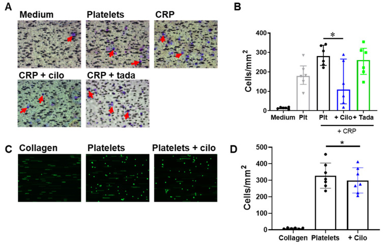
PDE3A inhibition decreases platelet-induced migration and platelet-induced adhesion of THP-1 cells under flow. Representative images (A) and quantitative analysis (B) of THP-1 cell migration to medium and to washed platelets, untreated or additionally stimulated with CRP and inhibited with cilostazol (5 μM) or tadalafil (10 nM). Red arrows indicate THP-1 cells. Representative images (C) or quantitative analysis (D) of THP-1 adhesion to collagen alone, and to washed platelets in the absence or presence of cilostazol (5 μM). Interquartile range (B), mean ± S.D. (D), n = 6 (A,B) or 7 (C,D), * p < 0.05. Statistics: Kruskal–Wallis test followed by Dunn’s multiple comparisons test (B) and Wilcoxon matched-pairs signed rank test (D). Cilo, cilostazol; CRP, collagen-related peptide; plt, platelets; tada, tadalafil.

