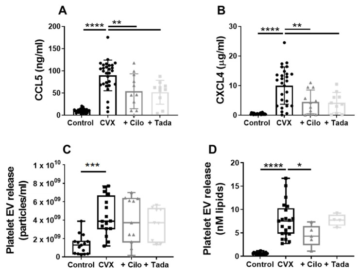
Figure 5.
Effect of PDE3A and -5 inhibition on convulxin-induced chemokine and extracellular vesicle release by platelets. Washed platelets were stimulated with convulxin (100 ng/mL) without or with cilostazol (5 µM) or tadalafil (10 nM), and the release of chemokines CCL5 (A) and CXCL4 (B), and of total (C) and procoagulant (D) platelet extracellular vesicle (EV) was measured. (A,B) Mean ± S.D., n = 27–29 (control, CVX) or 10–11 (cilo, tada). (C,D) Interquartile range, n = 15–16 (control, CVX; NTA), 9–11 (cilo, tada; NTA), 21–22 (control, CVX; PTase), 4–5 (cilo, tada; PTase), * p < 0.05, ** p < 0.01, *** p < 0.001, **** p < 0.0001. Statistics: ordinary one-way ANOVA followed by Dunnett’s (A,B) or Holm-Sidak’s (C,D) multiple comparisons test. Cilo, cilostazol; CVX, convulxin; NTA, nanoparticle tracking analysis; PTase, prothrombinase; tada, tadalafil.

